Research
Follow our publications on Dr. Stark’s google scholar, and also make sure to check out the free texts of all publications in our resources & paper copies tab
We often use technology and videoconferencing for our research. Here’s a short talk by Dr. Stark about some of this and a free copy of the paper including some of our best practices. See the Consulting page if you’d like to work with Dr. Stark on designing or visualizing a remote design when conducting research with persons with aphasia!
Research Topics

Overview
The lab evaluates how aphasia impacts spoken discourse (language beyond the sentence level) and, more globally, communication, via three inter-related focus areas: (1) characterizing language produced during spoken discourse using neuropsychological and neuroimaging methods; (2) identifying the extent to which spoken discourse is supplemented by gesture; and (3) examining how preserved inner speech (our inner narrative) supports aphasia recovery. We also have a longstanding interest in identifying the extent to which technology can improve assessment and intervention for persons with language disorders, especially aphasia. This research increases understanding of the fundamental aspects of spoken language and communication from an interdisciplinary perspective by leveraging theory and methodology derived from neuroscience, linguistics, communication sciences and disorders, and cognitive science. Our research is translational, having the potential to improve aphasia management by better understanding prognostic factors (e.g., brain damage associated with language impairments, inner speech’s role in predicting language recovery) and strategies that improve communication (e.g., use of gesture).
Click the link for each research topic to be taken to a page with more information, as well as relevant publications and multimedia presentations from our lab on the topic.
Inner Speech
I’d like to read about the technical details
I’d like to read the accessible summary
Inner speech is the internal dialogue (or, monologue) we engage in with ourselves. “Inner speech” has been called a variety of things: inner (or internal) monologue, inner (or internal) dialogue, inner voice, covert (or silent) self-talk, internal narrative, verbal thinking, endophasia, autocommunication, and so on. We prefer the term 'inner speech' because it emphasizes the active nature of the phenomenon and does not presume about features, e.g., whether it is a monologue or dialogue. Interestingly, some of the population does not experience inner speech and those that do tend to vary in how often they engage with inner speech, and for what purposes. Inner speech has long been studied through interdisciplinary lenses (philosophy, neuroscience, linguistics, psychology), and therefore many methods and designs have been used to explore it. While it has most typically been explored in cognitively healthy young adults, our lab has a particular interest in studying inner speech in persons with post-stroke aphasia, a language disorder, because it may be intact compared to aloud speaking or even comprehension abilities.
Spoken Discourse
I’d like to read about the technical details
I’d like to read the accessible summary
Spoken discourse is language used for a specific purpose, and is speech that goes beyond the sentence level. For example, you might tell a story about what you did last night — that’s spoken discourse! The goal of this research line is to integrate spoken discourse analysis into typical language assessment because, at present, analyzing discourse is not standard in clinical or research. the bulk of language and communication assessment in aphasia. Analyzing discourse enables simultaneous investigation of language structure (phonology, morphology, syntax, semantics, and pragmatics), dimensions of use (form, content, context), and multimodal strategies (gesture, eye gaze), lending a robust and comprehensive view of language and communication. During discourse, individuals with aphasia are enabled to speak on topics that are important to them. This establishes an inclusive and salient means of understanding communication ability. Finally, analyzing discourse may be a more sensitive means of establishing communicative competence across the aphasia severity spectrum. For example, discourse assessment identifies residual, subtle language impairments in the mildest forms of aphasia and then provides critical evidence for the receipt of continued clinical services to these individuals, many of whom want to rejoin the workforce. Our lab is very interested in how context and retelling affects discourse, and how discourse best practices (analysis, acquisition) can be improved.

Common genres in monologic discourse
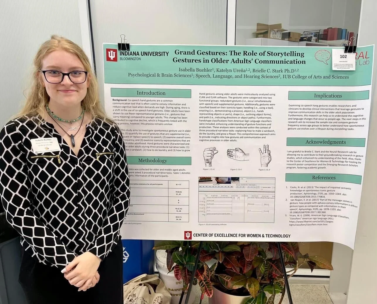
Communicative Gesture
I’d like to read about the technical details
I’d like to read the accessible summary
The short-term goal of this line of research is to understand how gestures improve communicative competence, which is the ability to successfully participate in the world using any verbal and non-verbal means. The long-term goal is to build assessments that enable quantification of gesture’s role in improving communication over time. Gesture appears to be remarkably intact in most individuals with aphasia, even in the presence of limb weakness or paralysis. We’re also collaborating with a computer scientist to see if an LLM-model can be trained to interpret speech and gesture from persons with aphasia, improving the current speech-to-text technology by creating a multimodal-to-text technology.
Brain Bases of Language and Language Recovery
I’d like to read about the technical details
I’d like to read the accessible summary
My clinical neuroscience research moves the neurobiology of language field forward by examining the neural mechanisms and structures related to impaired language processes during discourse, and prior research has focused on evaluating brain bases of language recovery in chronic aphasia. Most of the research used to establish a neurobiology of language evaluated relationships between brain damage and impaired, isolated language processes (e.g., naming). Discourse is important to evaluate because the cognitive components required for lexical retrieval in this environment are more complex than in the isolated environment: future and past lexical alternatives must be suppressed during discourse, while other processes, like syntax, occur in parallel. Therefore, by evaluating discourse impairments and their relationship to brain damage, my research provides complementary and novel evidence for theory-building in the neurobiology of language. I typically utilize structural and functional MRI to investigate brain bases of language impairment and recovery.
Technology & Aphasia
I’d like to read about the technical details
I’d like to read the accessible summary
Technology is a powerful tool to improve assessment and treatment of acquired language disorders, such as aphasia. With the advent of videoconferencing and remote technology (e.g., smartphone apps, immersive virtual reality) comes considerable opportunities, including increasing the ecological validity of assessment, upping treatment dosage, and demonstrating treatment generalization to real world contexts. I have been evaluating the intersection of technology and its use in assessment and intervention for aphasia since 2012, publishing one of the earliest clinical trial investigations of tablet-delivered speech therapy. The lab published a technical report on our pipeline for conducting language and cognitive assessment and intervention remotely and, since 2020, we have recruited >250 individuals with aphasia from across the USA to participate in our remote studies. We’ve also dabbled in using smartphones to measure behavior (see the Inner Speech page!) and Dr. Stark completed a Fulbright Scholar Award at the University of Technology Sydney in Spring 2025, where she collaborated on a pilot study of immersive virtual reality (those cool headsets!) to evaluate its potential use in persons with acquired language disorders. Presently, we are focusing on: (1) with University of Technology Sydney: studying how immersive virtual reality impacts story retelling and evaluating user experience of immersive virtual reality in persons with stroke and aphasia; (2) with University of South Florida: building and evaluating a gesture-sensitive LLM-based tool for enhancing communicative effectiveness in aphasia; and (3) with University of Maryland Global Campus and fellow Fulbrighter Jason Pittman, building and evaluating machine learning and LLM-based models for improving how we analyze discourse in aphasia.
Scroll down to learn about how we’re funded, our equipment and facilities, and a sampling of student research!
Funding
Dr. Stark’s research has been supported by the Gates Cambridge Trust, Indiana clinical and translational sciences institute, Indiana University, Indiana University Institute for Advanced Study, National Institute on Disability and Rehabilitation Research Switzer Merit Fellowship, the National Institute on Deafness and Other Communication Disorders, the ASHFoundation New Investigator Award, the National Aphasia Association Barbara Martin Research Award, the National Academy of Medicine Catalyst Award, and the Fulbright Scholar Award.
Press release from ASHFoundation HERE

Research Facilities
We are located within Indiana University’s Department of Speech, Language and Hearing Science
We work closely with Clinical Faculty in IU’s Speech-Hearing Clinic
Address:
2631 E Discovery Parkway
Bloomington, IN 47408

We collaborate closely with faculty and research scientists at Indiana University’s Psychological and Brain Sciences (PBS) Department
PBS houses our MRI facilities (Imaging Research Facility), including a 3T Siemens Prisma MR scanner
Address:
1101 E 10th St
Bloomington, IN 47405